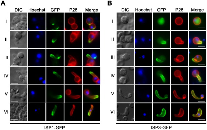
Fig. 3. ISP1 and ISP3 define apical polarity during zygote (ookinete) development and differentiation.
Localisation of ISP1-GFP (A) and ISP3-GFP (B) throughout the different stages of ookinete development (I–VI). A Cy3-conjugated antibody to P28 defines the surface of the zygote/ookinete, nuclei were detected using Hoechst dye, and the cells were displayed by differential interference contrast (DIC). Merge is the composite of Hoechst, GFP and P28. Scale bar = 5 µm.

