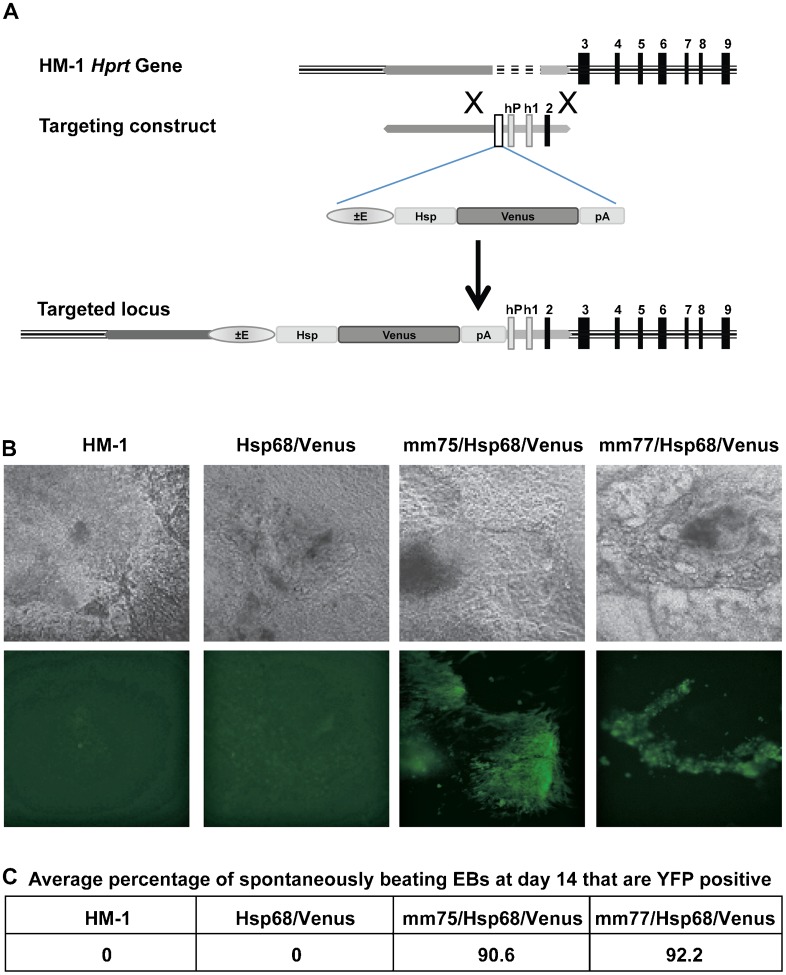
Fig. 1. Hprt targeting strategy and cardiac enhancer activity in beating cardiomyocytes.
(A) The Hprt locus in mouse HM-1 ES cells lacks the Hprt promoter, exon 1 and exon 2. The Hprt targeting vector contains the enhancer of interest, the Hsp68 minimal promoter and Venus fluorescent reporter gene upstream of the human HPRT promoter, human exon 1 and mouse exon 2 and these are all flanked by Hprt locus homology arms. Homologous recombination of this targeting construct in HM-1 ES cells results in reconstitution of the Hprt locus and insertion of the enhancer/Hsp68/Venus reporter upstream of the promoter. Use of Hprt substrate analogues to select for HM-1 with deficient or reconstituted Hprt provides a stringent selection method for selection of correctly targeted clones. (B) Bright field (above) and fluorescent (below) images of representative beating embryoid bodies (EBs) at day 14, from HM-1, Hsp68/Venus control, mm75/Hsp68/Venus and mm77/Hsp68/Venus clones. (C) Average percentage of spontaneously beating EBs that are YFP positive at day 14 for the above clones, average of two independent differentiation experiments.

