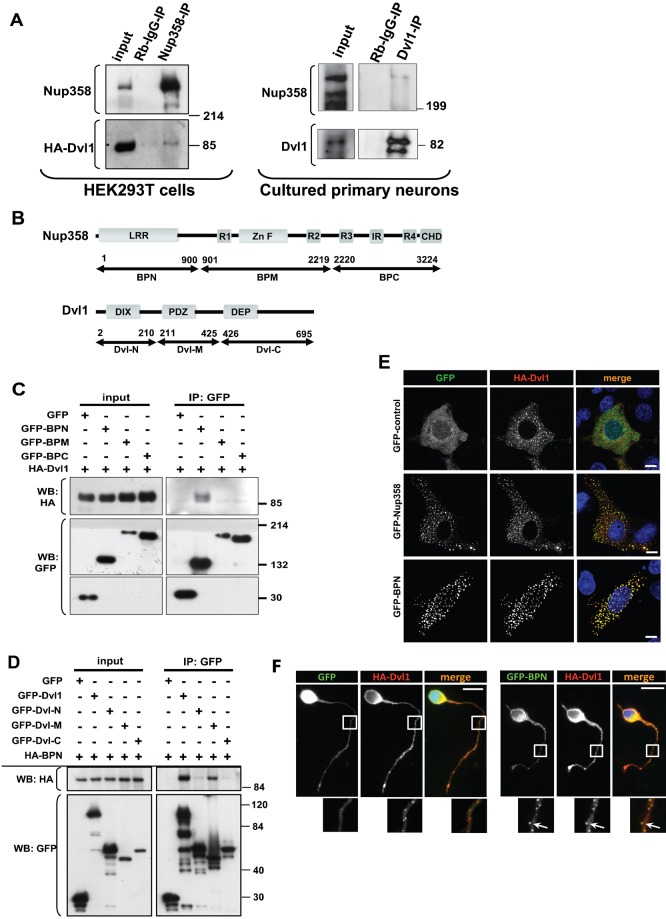
Fig. 1. Nup358 interacts with Dishevelled.
(A) Left panel: HA-tagged Dvl1 was overexpressed in HEK293T cells and endogenous Nup358 was immunoprecipitated (IP) using rabbit polyclonal antibodies against Nup358 (Nup358-IP). Rabbit IgG (Rb-IgG-IP) was used as control. Immunoprecipitates were analysed by western blotting (WB) with antibodies against HA and mAb414 (mouse monoclonal antibody that recognizes nucleoporins possessing FxFG sequences such as Nup358). Molecular weight markers are as indicated (in kDa). Right panel: Cultured primary neurons isolated from E18 rat were subjected to immunoprecipitation using Dvl1 antibody. The immunoprecipitate was probed with Dvl1 and Nup358 antibodies. (B) Domain structure of full-length Nup358 and Dvl1 and their fragments used in the study. The numbers refer to the amino acid positions. (C) HEK293T cells co-expressing HA-Dvl1 and GFP-control or GFP-tagged fragments of Nup358 were subjected to immunoprecipitation using anti-GFP antibodies. The immunoprecipitates were probed with antibodies against GFP or HA. (D) GFP-tagged full-length or fragments of Dvl1 were co-expressed with HA-BPN and were immunoprecipitated using anti-GFP antibodies. The immunoprecipitates were subjected to western analysis using indicated antibodies. (E) COS-7 cells were co-transfected with HA-Dvl1 (red) and GFP control, GFP-Nup358 or GFP-BPN (green) and were immunostained with anti-HA antibodies. DNA was visualized by Hoechst 33342 staining (blue). Scale bar, 10 µm. (F) E18 rat hippocampal neurons were transfected with pBetaActin-eGFP (GFP, green) or pBetaActin-BPN-eGFP (GFP-BPN, green) and pBetaActin-HA-Dvl1 (HA-Dvl1, red) constructs for 72 hours. The cells were fixed, stained and analyzed by fluorescence microscopy. DNA was stained with Hoechst 33342 dye. Scale bar, 20 µm. Arrows indicate co-localization of GFP-BPN with HA-Dvl1 puncta in neuronal extensions.

