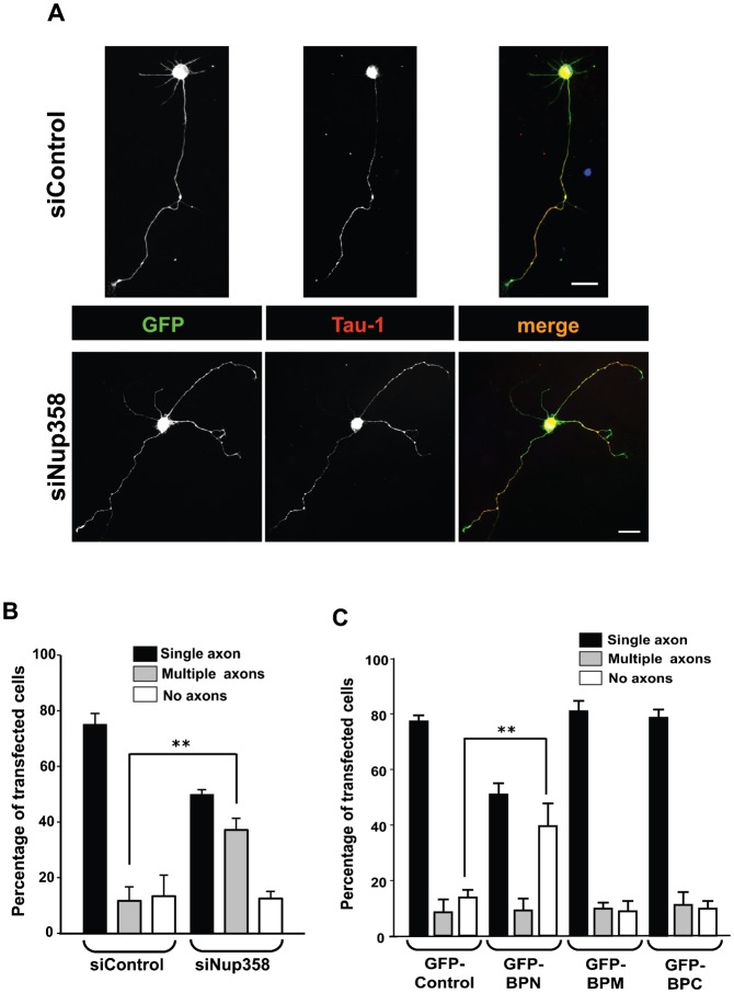
Fig. 4. Nup358 is required for neuronal polarization.
(A) E18 hippocampal neurons were transfected with control (siControl) or Nup358 siRNA (siNup358) along with pBetaActin-eGFP as transfection control, and were immunostained after culturing for 72 hours in vitro. The transfected neurons were identified by GFP expression (green). The effect of Nup358 depletion on axon formation was analysed using anti-Tau-1 antibodies (red). Arrows indicate axons as identified by Tau-1 axonal marker. DNA was stained with Hoechst 33342 dye (blue). Scale bar, 25 µm. (B) Quantitative analysis of the effect of Nup358 depletion on neuronal polarity. Error bars indicate standard deviations, n = 3, **P<0.01, Student's t test. (C) E18 rat hippocampal neurons were transfected with GFP-control, GFP tagged version of BPN, BPM or BPC and assessed for the effect on neuronal polarization after 72 hours. Error bars indicate standard deviations, n = 3, **P<0.01, Student's t test.

