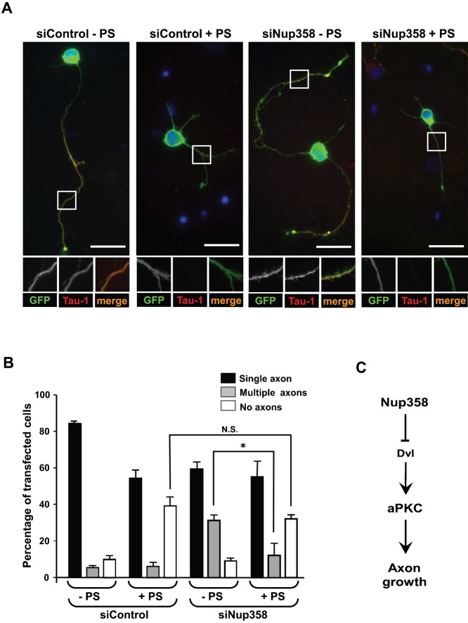
Fig. 6. Multiple axons formed by Nup358 depletion is partially rescued by inhibition of aPKC activity.
(A) Hippocampal neurons transfected with control (siControl) or Nup358 (siNup358) siRNA along with pBetaActin-eGFP and were cultured in the presence (+ PS) or absence (− PS) a pseudosubstrate inhibitor specific for PKCζ (10 µM). The neurons were fixed and immunostained after 72 hours. The effect of different treatments on neuronal polarization was assessed by scoring for GFP expression (green) and Tau-1 staining (red). Scale bar, 25 µm. (B) The quantitative analysis of the effect on neuronal polarity. Error bars indicate standard deviations, n = 3, *P<0.05, Student's t test. (C) A working model for the function of Nup358 in neuronal polarization. During the initial stages of single axon formation, Dvl-mediated activation of aPKC occurs at the nascent axon. Differentiation of other neurites into axons could be spatially and temporally inhibited by Nup358-mediated interference of Dvl and aPKC functions.

