Figure 2.

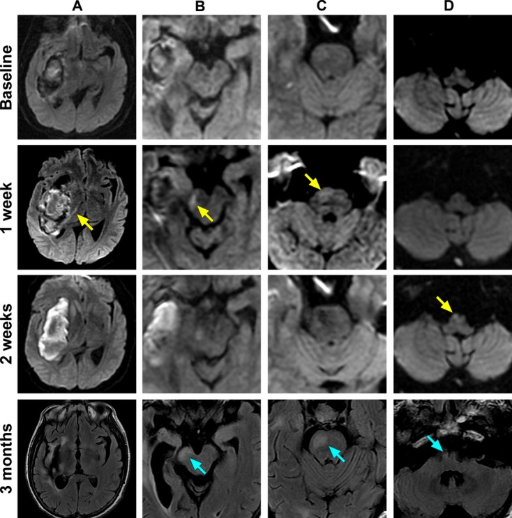
Spatial and temporal evolution of Wallerian degeneration in a single patient. Serial diffusion‐weighted imaging magnetic resonance images (MRIs) are shown for a single patient with a putaminal hemorrhage, obtained 2 days, 1 week, and 2 weeks after symptom onset. Corresponding fluid attenuated inversion recovery (FLAIR) sequences are shown at 3 months. A, Internal capsule. B, Cerebral peduncle. C, Pons. D, Medulla. Restricted diffusion appears along the ipsilateral corticospinal tract at 1 week in the posterior limb of the internal capsule, cerebral peduncle, and pons (arrows). At 2 weeks restricted diffusion appears in the medullary pyramid (arrow). At 3 months, areas that previously showed restricted diffusion are hyperintense on FLAIR and have undergone atrophy (arrows).
