
Figure 1.
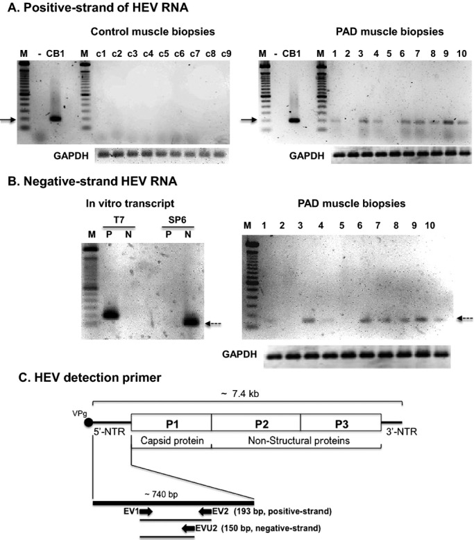
HEV RNA is present in muscle biopsies of PAD gastrocnemius. Total RNA from muscle homogenates of 9 controls and 10 PAD patients comprising those with disease Stage II (samples 2, 5, 7, and 9), Stage III (samples 1, 4, and 10) and Stage IV (samples 3, 6, and 8) were analyzed by RT‐PCR (T7 RNA polymerase) for HEV RNA (positive‐strand genomic RNA) (A). All controls had no detectable HEV RNA (left panel) while 8 of the 10 PAD specimens were positive for HEV RNA (right panel). RNAs of the 10 PAD patients also were analyzed by RT‐PCR (SP6 RNA polymerase) for negative‐strand viral RNA (indicating viral replication) (B). All PAD specimens that were positive for HEV RNA (specimens 1, 3, 4, 6 to 10) had detectable negative‐strand RNA and those PAD specimens negative for HEV RNA (2 and 5) had no detectable negative‐strand RNA. For a control, T7 RNA polymerase‐transcribed positive‐strand HEV RNA (P) and SP6 RNA polymerase‐transcribed negative‐strand viral RNA (SP6) were synthesized from a linearized pGEM‐T harboring CVB1EV1/2 fragment (data not shown). A 100 bp ladder is seen in lane “M”. GAPDH was used as an internal housekeeping gene. Solid arrows indicate 200 bp DNA and dashed arrows indicate negative‐strand viral RNA. (C), HEV RNA detection primers, EV1, EV2 and EVU2 are shown. The EV1/EV2 primer pair was used for detection of positive‐strand HEV RNA (193 bp). The EV1/EVU2 primer pair was used for detection of negative‐strand viral RNA (150 bp). HEV indicates human enterovirus; PAD, peripheral arterial disease; GAPDH, glyceraldehyde 3‐phosphate dehydrogenase; RT‐PCR, reverse‐transcription polymerase chain reaction; VP1, viral capsid protein.