
Figure 6.
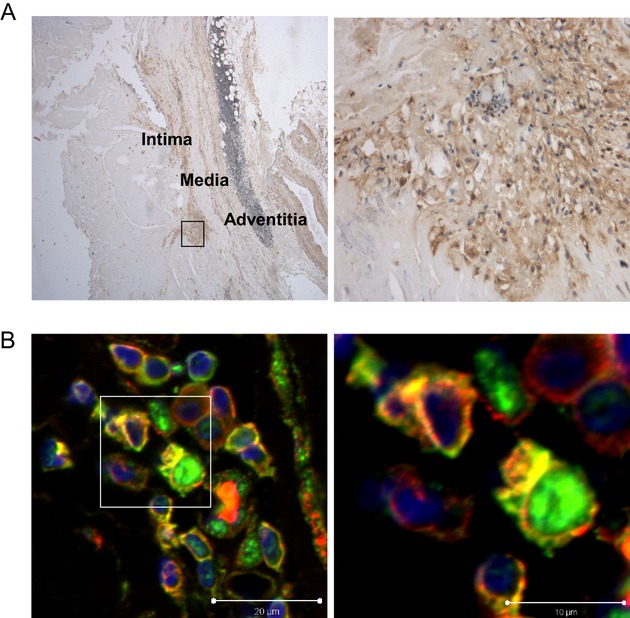
Rev‐erbα protein is present in the macrophages of human carotid artery atherosclerotic lesions. Human carotid artery sections were subjected to immunohistochemistry using mouse monoclonal anti‐human Rev‐erbα. A, Rev‐erbα immunoreactivity in a representative plaque (40×) (left panels), and a high‐magnification view of the same image (400×) (right panels) illustrating the cellular nature of the staining. B, Immunofluoresence staining identifying the Rev‐erbα positive cells as macrophages. A representative section of a human carotid plaque stained by using primary antibodies against Rev‐erbα (green) and the macrophage‐specific marker CD68 (red). Colocalization of the two markers is shown in yellow (left panels), and a higher magnification view of the same immunofluorescence image (right panels).