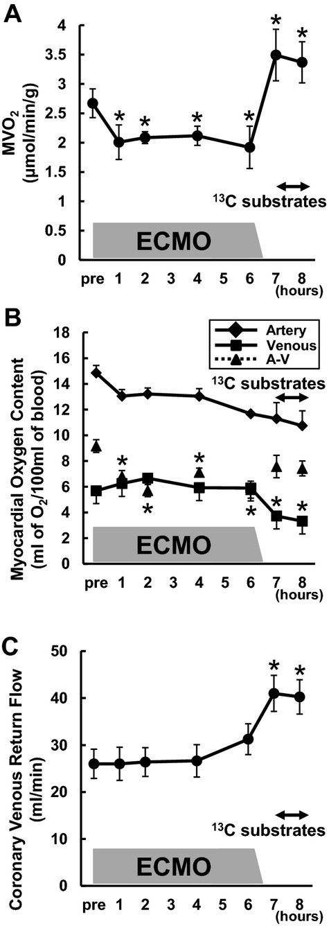
Figure 2.

Myocardial oxygen consumption rate (MVO2) in RELOAD. The time‐course of changes in MVO2 (A), oxygen content in the coronary vessels (B), and coronary flow (C). MVO2 was markedly decreased with ventricular unloading (*P<0.05 vs baseline), and increased more than baseline by RELOAD (*P<0.05 vs baseline). Substrate infusion itself did not lead to change MVO2. Moreover difference between arterial and venous oxygen content was significantly decreased during ECMO, whereas coronary flow was significantly increased by RELOAD (*P<0.05 vs baseline). Values are means±SE; n=8. ECMO indicates extracorporeal membrane oxygenation; SE, standard error; A‐V, difference between arterial and venous oxygen content.
