
Figure 3.
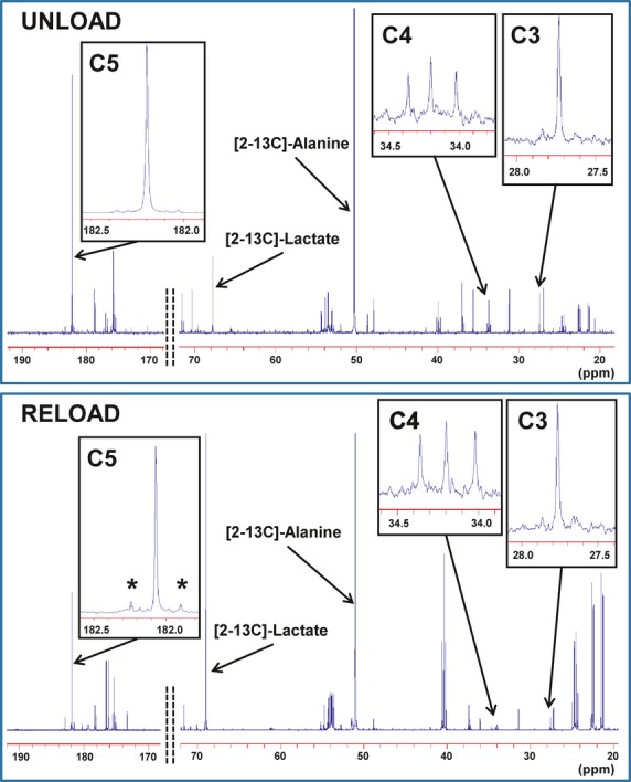
Typical 13C‐NMR spectra obtained from left ventricular extract after a 60‐minute infusion of [2‐13C]‐pyruvate and [U‐13C]‐leucine into the left anterior descending coronary artery under UNLOAD and RELOAD conditions. The spectra show adequate signal to noise ratio for peak integration. Chemical shifts in parts per million (ppm) were as follows: C3, 27.7; C4, 34.2; C5 of glutamate, 182.2; [2‐13C]‐alanine, 51.9 and [2‐13C] lactate, 69.2. Marked differences occur in glutamate peak complexes between the 2 conditions. C5 in RELOAD shows prominently increased doublet peak area (*), indicating increased leucine contribution or decreased pyruvate contribution, compared to UNLOAD. Additionally, the greater labeled lactate to alanine ratio can be observed in RELOAD. NMR inidcates nuclear magnetic resonance.