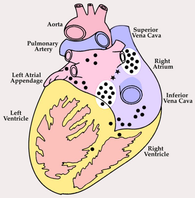
Figure 1.

Posterior view of adult human heart showing distribution of cardiac ganglia on the surface (•) and in the interatrial septum (*). Cardiac ganglia adjacent to the sinoatrial and atrioventricular nodes (white ovals) were harvested for morphological studies from normal human hearts and those with ischemic heart failure; homologous regions from canines with ischemic heart failure and spontaneously hypertensive rats with heart failure secondary to hypertension were also harvested. Modified from Singh et al.32
