
Figure 4.
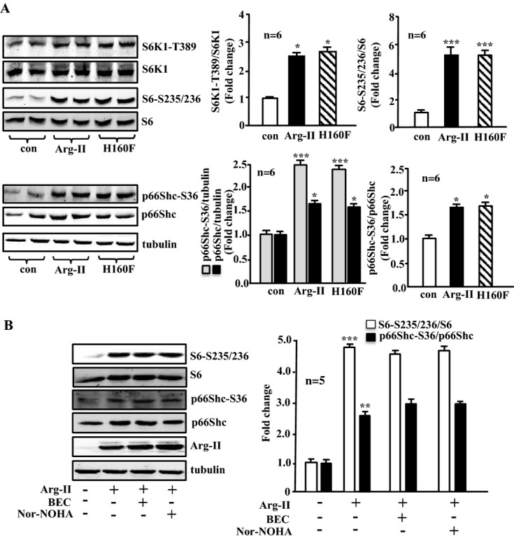
Overexpression of Arg‐II in young VSMCs activates S6K1 and p66Shc independently of its enzymatic activity. A, Young VSMCs were transduced as in Figure 1. Shown is immunoblotting analysis of phosphorylated S6K1‐T389 and total S6K1, phosphorylated S6‐S235/236 and total S6, and phosphorylated p66Shc‐S36 and total p66Shc levels. Tubulin served as a loading control. B, Young VSMCs were transduced with empty vector rAd/CMV as control (Arg‐II: −) or rAd/CMV‐Arg‐II (Arg‐II: +). Cells were treated with or without the arginase inhibitor BEC (200 μmol/L) or nor‐NOHA (50 μmol/L) overnight during serum starvation before cell lysate preparation 72 hours posttransduction. Shown is the immunoblotting analysis of phosphorylated S6‐S235/236 and total S6, phosphorylated p66Shc‐S36, and total p66Shc levels and Arg‐II level. Tubulin served as a loading control. Bar graphs show quantifications of the signals (n=6 in A, n=5 in B). *P<0.05, **P<0.01, ***P<0.001 vs control. Arg‐II indicates arginase‐II; con, control; VSMC, vascular smooth muscle cell; rAd, recombinant adenovirus; BEC, S‐12‐bromoethyl‐l‐cystine; Nor‐NOHA, Nω‐hydroxy‐nor‐l‐arginine.