
Figure 8.
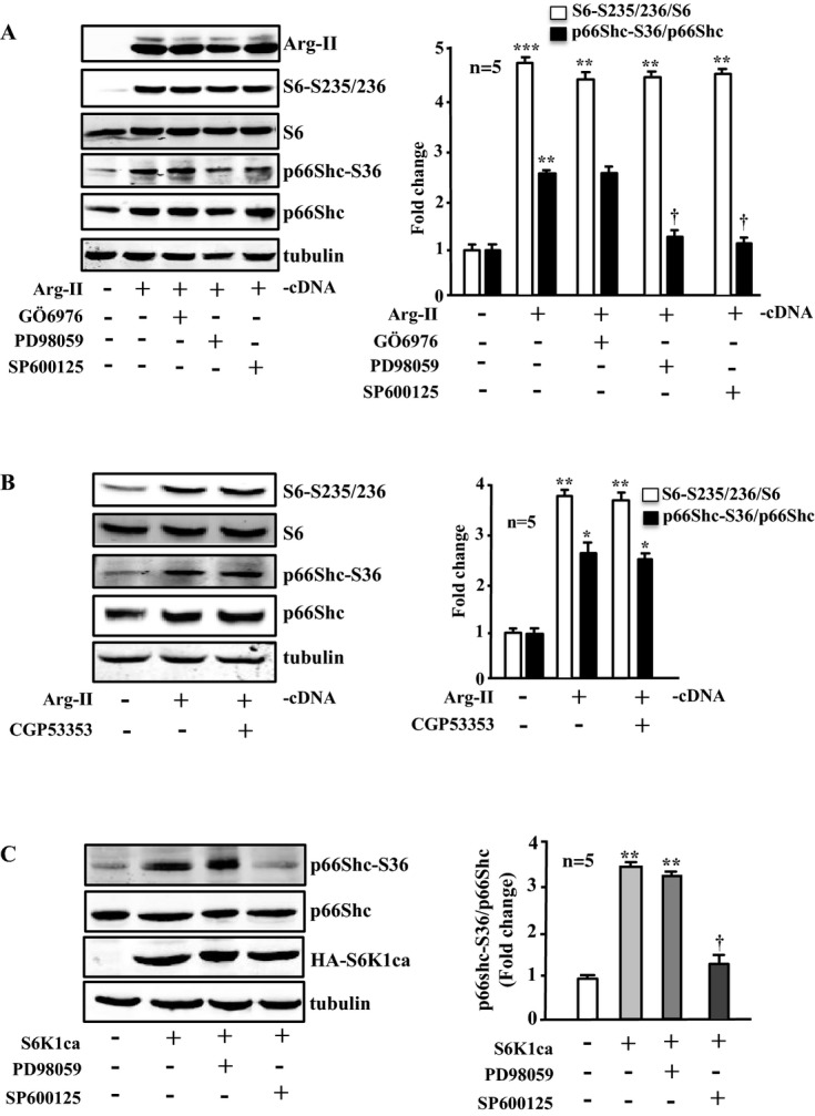
Effects of signaling inhibitors on Arg‐II‐ or S6K1‐induced phosphorylation of p66Shc‐S36. Immunoblotting analysis of the signaling and protein levels as indicated. A and B, Young VSMCs were transduced with empty‐vector rAd/CMV as control (Arg‐II: −) or rAd/CMV‐Arg‐II (Arg‐II: +). Cells were treated with or without the signaling inhibitor overnight during serum starvation before cell lysate preparation 72 hours posttransduction. Cells were treated with Gö6976 (1 μmol/L), PD98059 (50 μmol/L), or SP600125 (20 μmol/L) or with CGP53353 (1 μmol/L) as indicated. C, Young VSMCs were transduced with empty‐vector rAd/CMV as control (S6K1ca: −) or rAd/CMV‐HA‐S6K1ca (an active mutant of S6K1; S6K1ca: +). Cells were treated with PD98059 or SP600125 as described in A and B. Quantification of the signals is shown in the corresponding bar graphs (n=5). *P<0.05, **P<0.01, ***P<0.001 vs control; †P<0.05 vs Arg‐II (in A) or vs S6K1ca (in C). Arg‐II indicates arginase‐II; VSMC, vascular smooth muscle cell; SA‐β‐gal, senescence‐associated β‐galactosidase; H2DCF, 2′,7′‐dichlorofluorescein; rAd, recombinant adenovirus.