
Figure 13.
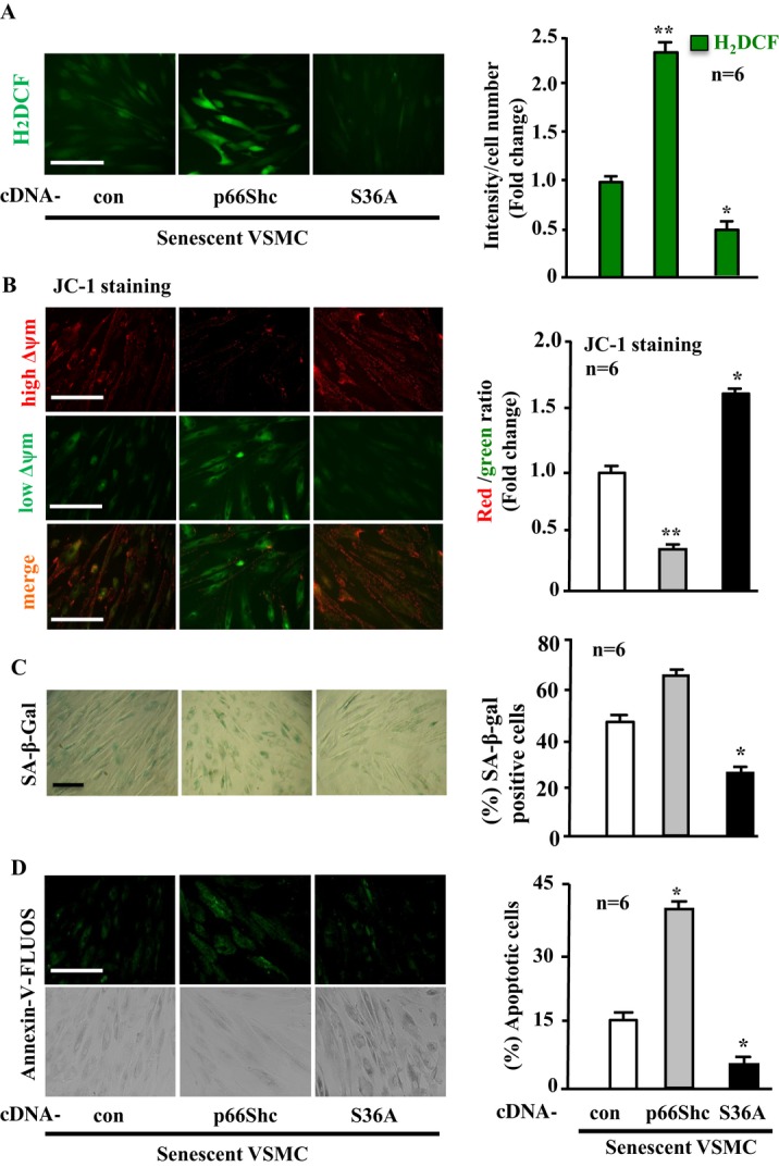
Enhanced phosphorylation of p66Shc‐S36 accounts for the phenotypic changes of senescent VSMCs. Senescent VSMCs were transduced with either rAd/CMV empty vector as control (con), rAd/CMV‐p66Shc or ‐p66Shc‐S36A (S36A) as indicated. Seventy‐two hours posttransduction, the cells were subjected to (A) H2DCF staining for detection of H2O2. B, JC‐1 staining for analysis of Δψm. C, SA‐β‐gal staining for senescent cells. D, Annexin‐V‐FLUOS staining for apoptotic cells. Quantification of the signals is shown in the corresponding bar graphs (n=6). *P<0.05, **P<0.01 vs con. Scale bar=0.2 mm. Arg‐II indicates arginase‐II; VSMC, vascular smooth muscle cell; H2DCF, 2′,7′‐dichlorofluorescein; SA‐β‐gal, senescence‐associated β‐galactosidase; rAd, recombinant adenovirus.