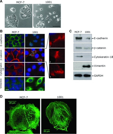
Fig 1.

Acquisition of cell resistance to TNF-α is accompanied by morphological changes and actin cytoskeleton reorganization. (A) The morphology of TNF-sensitive MCF-7 and TNF-resistant 1001 cells by phase contrast microscopy. Bar: 100 μm. (B) Immunofluorescence analysis of epithelial and mesenchymal markers. Cells were labelled with E-cadherin, β-catenin, cytokeratin-18 or vimentin primary antibody. Secondary antibodies were Alexa-Fluor 488-conjugated goat antimouse IgG for E-cadherin and vimentin (green) and Alexa-Fluor-594-conjugated goat antimouse IgG for β-catenin and cytokeratin-18 (red). Nuclei were stained with DAPI (blue). Cells were analysed by epifluorescence microscopy (LeicaDMRX microscope). Three enlarged regions of β-catenin staining in 1001 cells are shown. Bar: 10 μm. (C) Expression of epithelial and mesenchymal marker proteins in MCF-7 and 1001 cells. Immunoblot analysis was performed on total protein extracts (50 μg) using E-cadherin-, β-catenin-, cytokeratin-18-, vimentin- or GAPDH-specific antibody as previously described [16]. (D) Actin cytoskeleton organization in MCF-7 and 1001 cells. Cells were stained with Alexa-Fluor 488-coupled phalloidin to visualize F-actin and analysed using a Zeiss laser scanning confocal microscope (LSM-510 Meta). Bar: 20 μm.
