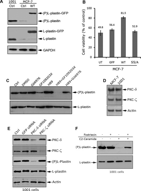
Fig 3.

Overexpression of phosphorylated L-plastin in MCF-7 cells confers resistance to TNF-α. (A) Ser5 phosphorylation level of endogenous L-plastin in control 1001 and MCF-7 cells (Ctrl) and L-plastin-GFP-transfected MCF-7 cells (WT). Immunoblot analysis was performed using an anti-ser5 phosphorylated L-plastin specific antibody (Ser5-P) [16] (upper panel), an anti-L-plastin (middle panel) or an anti-GAPDH (lower panel). The higher band (upper and middle panels) corresponds to the transfected L-plastin fused to GFP (L-plastin-GFP) and the lower band corresponds to endogenous L-plastin (L-plastin). (B) Unphosphorylable L-plastin does not confer resistance to TNF-α in MCF-7 cells. Cell viability assay was performed after 72 hrs of TNF-α treatment (75 ng/ml) on untransfected (UT) and transfected MCF-7 cells with either GFP (GFP), WT L-plastin-GFP (WT) or unphosphorylable L-plastin-GFP (S5/A). The transfection efficiency was nearly identical in cells (data not shown). Results are expressed as the mean ± S.D. of three independent experiments. (C) High baseline phosphorylation of L-plastin in 1001 cells involves non-conventional PKC isoforms. Cells were treated for 3 hrs with vehicle (DMSO), 0.5 μM of Gö6976 (conventional PKC inhibitor), 5 μM of GF109203X (conventional and non-conventional-PKC inhibitor), 5 μM of H-89 (PKA inhibitor). Cells were also treated with H-89 (5 μM) combined to either GF109203X (5 μM) or Gö6976 (0.5 μM).Immunoblot analysis was performed using anti-Ser5-P (upper panel) or anti-L-plastin (lower panel) antibody. (D) Expression of PKC-δ and –ζ in MCF-7 and 1001 cells. Immunoblot analysis was performed on total protein extracts (50 μg) using an anti-PKC-δ (Upper panel), an anti-PKC-ζ (middle panel), or an anti-actin (lower panel) antibody. (E) Knockdown of PKC-δ decreased L-plastin phosphorylation in 1001 cells. 1001 cells were transfected with either PKC-δ or PKC-ζ siRNAs (100 pmol) as well as with GFP siRNA used as a control. The protein expression levels of PKC-δ and ζ as well as L-plastin and phosphor-L-plastin were assessed by immunoblot in control untransfected cells (Ctrl), GFP-, PKC-δ or PKC-ζ siRNA transfected 1001 cells. The anti-actin antibody was used as a loading control. (F) Exogenous cell permeable C2-ceramide induces a decrease in L-plastin phosphorylation in 1001 cells by a mechanism involving the activity of the PP2A. Cells were untreated (–) or pre-treated (+) with 100 nM of Fostriecin. Cells were further incubated in the absence (–) or presence (+) of 10 μM C2-ceramide for 45 min. Immunoblot analysis was performed using anti-Ser5-P (upper panel) or anti-L-plastin (lower panel) antibody.
