
Fig 4.
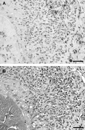
Identification of CD3-positive cells. (A) Immunohistochemistry of mesenteric polyarteritis lesions with anti-CD3 (DAB staining). Positively stained cells are labelled brown. (B) Chloracetate esterase reaction of mesenteric polyarteritis lesions for the identification of polymorphic leucocytes (red). Bar = 100 μm.