
Fig. 7.
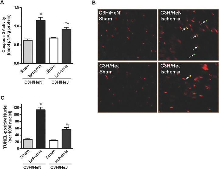
Myocardial apoptotic activity following in vivo regional ischemia. (A) Caspase-3 activity in hearts from C3H/HeN and C3H/HeJ mice. (B) Representative composite confocal photomicrographs of sections from C3H/HeN and C3H/HeJ hearts following ischemia. Hearts underwent TUNEL staining with fluorescein-labelled dUTP and counterstaining with propidium iodide. TUNEL+ nuclei are stained yellow (arrows). (C) The graph shows the quantification of apoptotic nuclei from C3H/HeN and C3H/HeJ hearts following ischemia. Values are mean ± S.E. for four experiments, *P< 0.05 versus control. †P < 0.05 versus C3H/HeN.