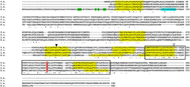
Figure 1.

Alignment of trypanosomatid mucolipin orthologues with human MCOLN1. The putative transmembrane regions (yellow), PI(3,5)P2 binding (green) or serine lipase active site (cyan) (LaPlante et al., 2011) are all highlighted. The boxed region represents the cation channel domain. The white triangle corresponds to a V→L alteration seen in MLIV patient 2 (Altarescu et al., 2002), while the black triangle relates to D→Y change that abolishes Fe2+ transport in mammalian cells (Dong et al., 2008). A transgene carrying the D→K mutation cannot complement MLIV cells (Pryor et al., 2006). Identifiers: T.b. – Trypanosoma brucei Tb927.7.950, T.c. –Trypanosoma cruzi TcCLB.508215.6, L.m. – Leishmania major LmjF.26.0990, H.s. – Homo sapiens NP_065394.1 (kinetoplastid genes identified with TriTrypDb/GeneDb codes, mammalian proteins with NCBI locus numbers). Asterisks indicate conserved residues. Alignment was carried out using clustalw2 at http://www.ebi.ac.uk/Tools/msa/clustalw2/.
