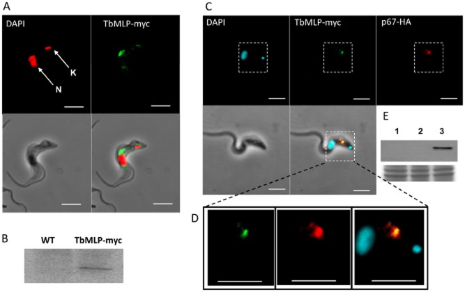
Figure 2.
Localization of carboxyl-terminally c-myc-tagged TbMLP (TbMLP-myc) in bloodstream-form trypanosomes.A. TbMLP-myc was localized with monoclonal antibody 9E10 and is shown as green; red indicates the nucleus (N) and kinetoplast (K) stained with DAPI. TbMLP is concentrated in a vesicular structure between nucleus and kinetoplast with some lighter staining around the flagellar pocket. White bar represents 5 μm.B. Western blot to show that antibody 9E10 recognizes a specific band in TbMLP-myc transformed cells; WT indicates wild type.C. TbMLP-myc colocalizes with lysosomal glycoprotein p67. To confirm lysosomal localization of TbMLP-myc the lysosomal marker protein p67 was also tagged with the HA epitope (p67-HA). TbMLP-myc localization is shown in green, p67-HA in red and DAPI stained DNA in cyan. White bar represents 5 μm.D. Magnification of the region in the dotted box to better show the colocalization of TbMLP-myc.E. Western blot to show the specificity of the anti-HA antibody for p67 (lane 1: wild type, 2: trypanosomes transformed with TbMLP-myc, 3: trypanosomes transformed with TbMLP-myc and p67-HA). The lower panel shows Coomassie-stained gel to show equivalent loading.

