
Fig 9.
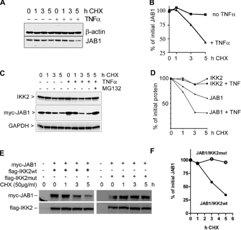
Degradation of JAB1. (A) Degradation of endogenous JAB1 is accelerated after treatment with TNFα as assessed by immunoblotting of 293 cell extracts prepared after timed addition of cycloheximide. (B) Quantification of the JAB1 band shown in (A). (C) Enhanced degradation of ectopically expressed JAB1 but not IKK2 after addition of TNFα. Myc-tagged JAB1 was expressed in 293 cells and protein neo-synthesis was blocked for different time periods by addition of cycloheximide. TNFα (50 ng/ml) was added to one part of the samples. Cell extracts were analysed by immunoblotting for IKK2 and myc-JAB1. (D) Quantification of the Western Blot bands shown in (C). E, degradation of myc-JAB1 in presence of either wild-type IKK2 (flag-IKK2wt) or kinase-deficient mutant IKK2 (flag-IKK2mut) after blocking protein synthesis for different time periods with cycloheximide (CHX). (F) quantification of the myc-JAB1 bands shown in (E).