
Fig 2.
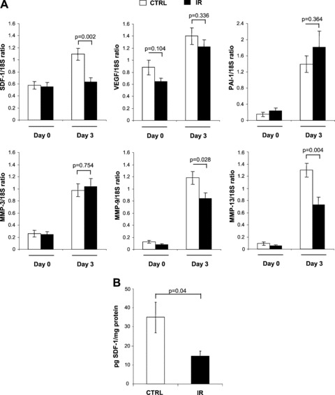
Absence of SDF-1α induction at the RNA and protein levels in wounded skin previously exposed to IR. (A) Total RNA was collected from skin (day 0) or from wounded tissues together with surrounding margins (day 3), and the ratios of SDF-1α, VEGF, PAI-1, MMP-3, MMP-9 and MMP-13 RNA relative to 18S ribosomal RNA were determined by quantitative real-time PCR. Shown is the expression levels as detected in mice previously exposed to IR (black bar) compared to control non-irradiated mice (white bar). (B) Amount of SDF-1α protein was determined by ELISA on wound lysates obtained from irradiated mice (black bar) compared to non-irradiated mice (white bar) 3 days after skin injury. Results are expressed as pg of SDF-1α protein per mg of total protein isolated per wound. Data are mean ± S.E.M.; (n= 9 mice per group, where each RNA and protein sample is composed of an average of two wounds per mouse).