
Fig 4.
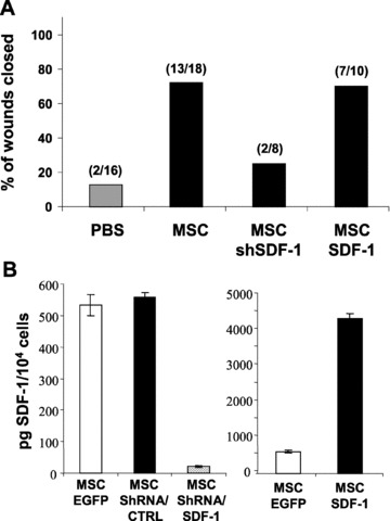
Specific secretion of SDF-1α by MSCs enhanced wound healing. (A) Full-thickness wound biopsies were created on the dorsal surface of mice previously exposed to IR and wound healing was determined following the intradermal injection (4 injections sites per wound) of purified populations of MSC, MSC-ShSDF-1, MSC-SDF-1 or PBS. As in Fig. 3, the proportions of wounds over 90% closed at day 13 are shown. (B) Amount of secreted SDF-1α in conditioned medium from MSCs genetically modified to express EGFP, shSDF-1, shCTR or SDF-1 was determined by ELISA. Data are expressed as means ± S.E.M. of three independent measurements.