
Fig 5.
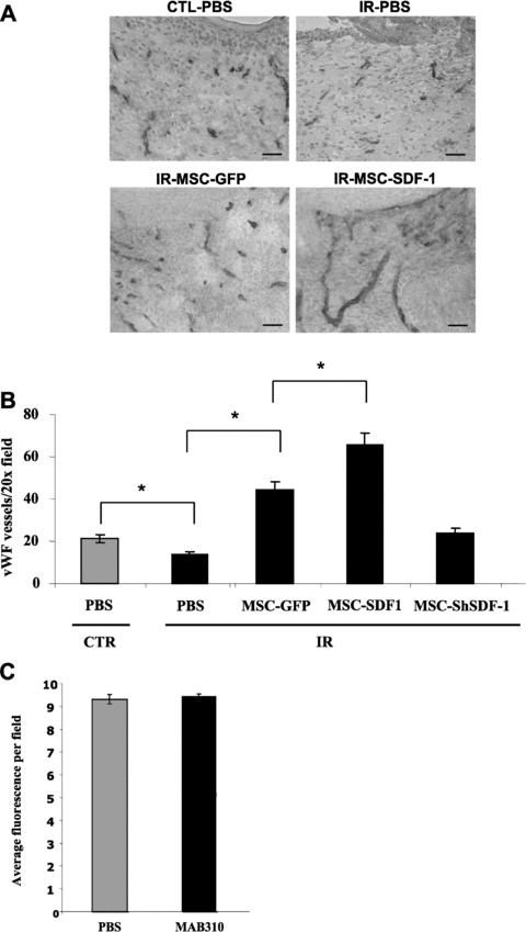
Increased wound vascularization by stromal cell injection. Full-thickness wound biopsies were created on the dorsal surface of mice, and purified populations of MSC-EGFP, MSC-ShSDF-1, MSC-SDF-1 in PBS or PBS alone were injected intradermally the following day (as described in Figs 3 and 4). Wound vascularization density, as determined by vWF stainings, was analysed 15 days after injury. Representative pictures of cryosections counterstained with haematoxylin are shown. Scale bar = 250 μm. (B) Quantification of the number of vWF positive vessels per 20× field for each group. Data are mean ± S.E.M.; (n= 6 to 12 sections from n= 3 to 5 mice; *P < 0.001). (C) Quantification of the proportion of GFP+ bone marrow-derived cells infiltrating the periphery of wounds injected daily with PBS or 40 μg of MAB310 (SDF-1α blocking antibody) at day 7 after injury (shown is the average of three photos per wound, each collected from n= 7 mice per group).