Fig 5.

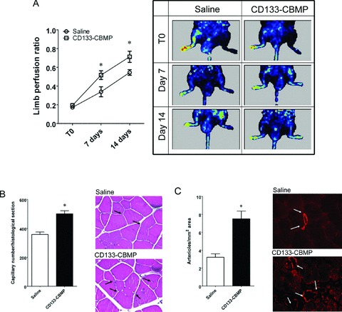
In vivo potency test of CB-derived CD133 CBMPs. (A) shows the increase in perfusion ratio in saline and CD133 CBMP-injected limbs. The LDPI imaging sequences show the increase in perfusion of the same saline-injected or CD133 CBMP-injected mice at the three times considered (T0, 7 and 14 days after removing the femoral artery). Plot shows the quantitative evaluation of the limb perfusion ratio in saline- (n= 8) and CD133 CBMP-(n= 11) injected mice at the three times considered. * indicates P < 0.05 by two ways unpaired t-test. (B) shows the capillary density in ischemic limbs injected with saline solution (n= 13) and CD133 CBMPs (n= 16). Insets show the capillaries in injected adductor muscles. * indicates P < 0.05 by two ways unpaired t-test. (C) shows the increase in the number of arterioles per histological section, as detected by smooth muscle actin staining in the same animals. Insets show the arterioles stained by α-actin antibody. * indicates P < 0.05 by two ways unpaired t-test.
