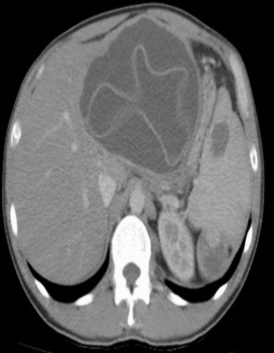
Fig. 1.

A 39-year-old male patient with splenic hydatid cyst disease. Axial CT sections show lesions consistent with hydatid cysts in the left lobe of the liver and in the spleen. A suspicious lesion of possible hydatid cyst origin is visible in the upper pole of the left kidney.
