FIGURE 3.

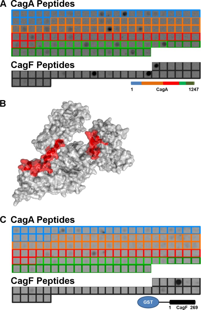
Interaction of recombinant CagA and CagF on CagA and CagF 15-mer peptide arrays. A, a peptide array consisting of CagA (blue, orange, red, light green, and dark green boxes denoting domains I, II, III, IV, and V respectively) and CagF (black boxes) peptides was probed for binding with GST-CagF and developed with GST antibody and HRP-anti-mouse IgG conjugate. B, the three most intense CagA peptides that bind GST-CagF (red) are mapped onto the structure of domains I–III of CagA (Protein Data Bank code 4DVZ). C, a peptide array consisting of CagA and CagF (same color schemes as described in A) was probed for binding with full-length CagA and developed with HRP-anti-His conjugate.
