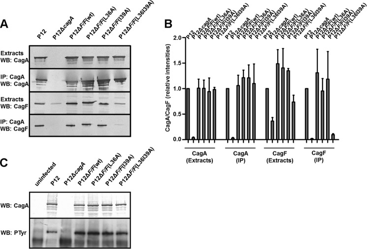
FIGURE 6.
Functional characterization of coiled coil CagF variants. A, whole-cell lysates of the indicated H. pylori strains were subjected to CagA immunoprecipitation. Extracts and IP fractions were analyzed by Western blot for CagA and CagF. B, densitometric quantification of CagA and CagF in IP experiments. Data are shown as means ± S.D. (error bars) for immunoblots obtained from at least three independent IP experiments. C, AGS cells were infected for 4 h with the indicated strains or left uninfected. Infection lysates were analyzed by Western blot (WB) with CagA- and phosphotyrosine (PTyr)-specific antibodies.

