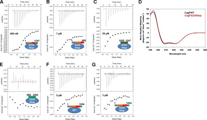
FIGURE 7.
The C-terminal helix of CagF folds upon binding CagA and subsequently interacts with domain IV of CagA. Isothermal titration calorimetry binding curves of the CagAWT-CagFS234Stop (A), CagA1–885-CagFS234Stop (B), and CagA1055–1247-CagFS234Stop (C) interactions are shown. D, comparison of the circular dichroism spectra of CagFWT (black line) and CagFS234Stop (red line) showing the latter to be more helical. Isothermal titration calorimetry binding curves of the CagA885–1054-CagFS234Stop (E), CagA1–1054-CagFWT (F), and CagA1–1054-CagFS234Stop (G) interactions are shown.

