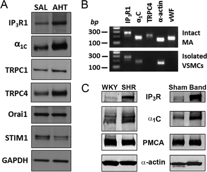
FIGURE 2.

Screening of Ca2+-handling proteins reveals coupled up-regulation of the vascular IP3R and LTCC in experimental models of hypertension. A, immunoblot analysis of IP3R1, α1C, TRPC1, TRPC4, Orai1, and STIM1 in lysates from mouse MA. MA lysate pooled from three SAL or AHT mice was added to each lane. Increased expression levels of IP3R1, α1C, and TRPC4 are detected in MA of AHT mice, whereas levels of TRPC1, Orai1, and STIM1 were similar. B, agarose gel analysis of products obtained after PCRs for IP3R1 (274 bp), α1C (185 bp), and TRPC4 (226 bp) using specific primers. α-Actin (152 bp) and vWF (178 bp) were used as VSM and endothelial cell markers, respectively. IP3R, α1C, and α-actin were detected both in intact MA and isolated VSM cells as expected. In contrast, TRPC4 and vWF were detected in arteries but not isolated VSM cells, implying expression only in endothelium. C, immunoblots comparing the expression of IP3R, α1C, PMCA, and α-actin (as a loading control) between MA from normotensive Wistar Kyoto rats (WKY) and SHR and between MA from sham-operated and aortic-banded (Band) hypertensive rats. In each case, lysates were pooled from arteries of three or four rats. Only the IP3R and α1C proteins are up-regulated in MA from hypertensive SHR and aortic-banded rats. Blots are representative of three experiments.
