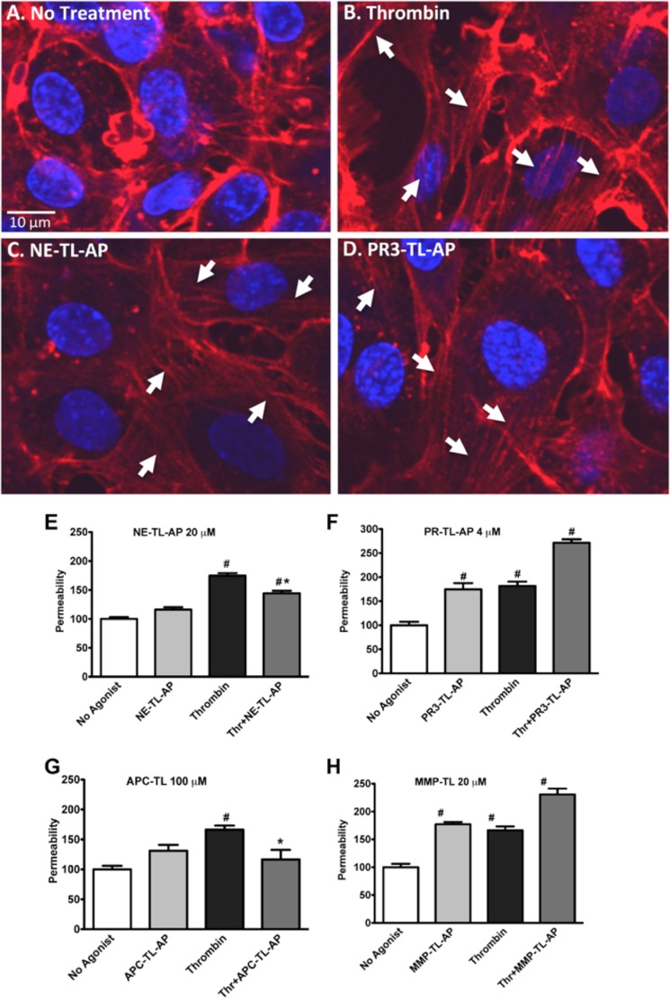
FIGURE 8.
Induction of F-actin stress fiber formation and modulation of permeability in HUVECs by NE-TL-AP and PR3-TL-AP. Confocal microscopy image of phalloidin-labeled F-actin (red) and DAPI-labeled nucleus (blue) in HUVECs treated with no agonist (A), thrombin (5 nm) (B), NE-TL-AP (20 μm) (C), and PR3-TL-AP (4 μm) (D). Integrity of the endothelial barrier was monitored by tracking the flux of FITC-dextran across HUVEC monolayers cultured on transwell supports following treatment with thrombin (5 nm) in combination with NE-TL-AP (20 μm) (E), PR3-TL-AP (4 μm) (F), APC-TL-AP (100 μm) (G), and MMP-TL-AP (20 μm) (H) as indicated. Permeability (% control) was normalized to the fluorescent reading obtained in the lower chamber for cells that had not been treated with agonists (expressed as 100%). Data are expressed as mean ± S.E. * indicates significant difference (p < 0.05) compared with agonist-untreated cells and # indicates significant difference (p < 0.05) compared with thrombin-treated cells (n = 6 for E and F; n = 3 for G and H).

