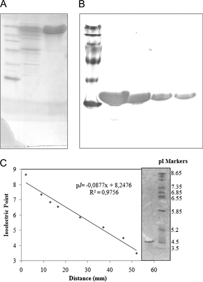
FIGURE 1.

Analysis of purified β-glucosidase from A. niger. A, SDS-PAGE of purified enzyme (1st lane, standard protein markers (14–116 kDa); 2nd lane, commercial preparation Novozymes 188; 3rd lane, purified β-glucosidase). B, native-PAGE of purified enzyme at different concentrations (10, 5, 2.5, and 1 mg/ml). C, isoelectric focusing of purified β-glucosidase (1st lane, standard pI markers; 2nd lane, purified β-glucosidase).
