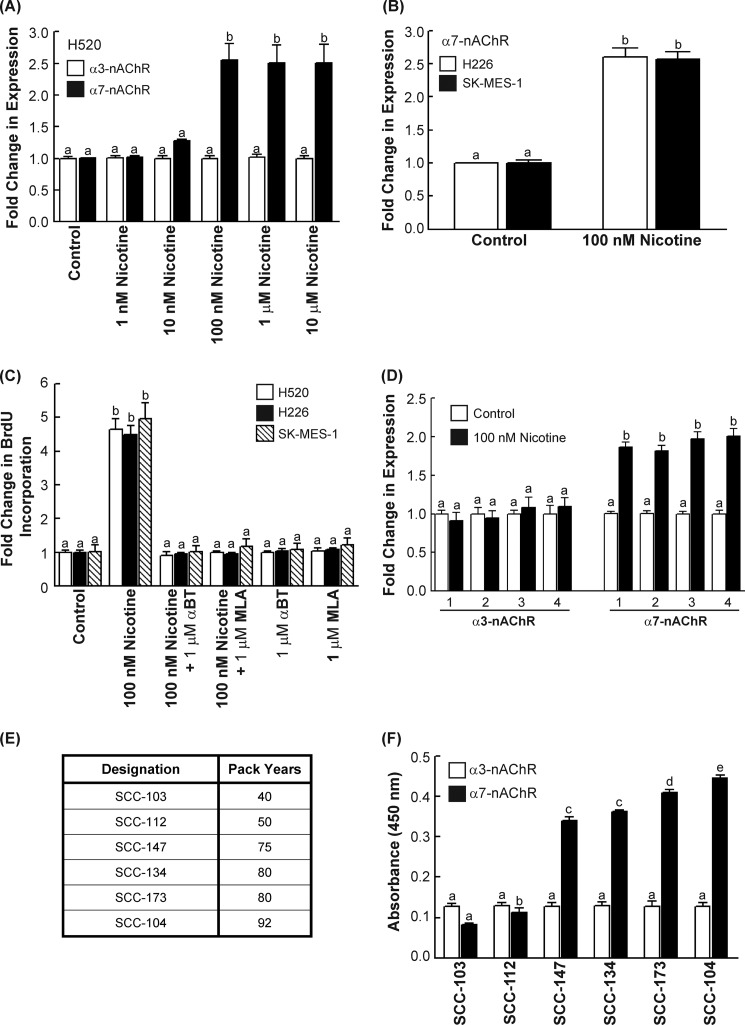
FIGURE 1.
Nicotine increased α7-nAChR expression in human SCC-L in vitro and in vivo. A, nicotine up-regulated the levels of α7-nAChRs (black bars) in a concentration-dependent manner in H520 human SCC-L cells in 96 h. However, nicotine did not affect the levels of α3-nAChRs (white bars). B, the treatment of H226 and SK-MES human SCC-Ls with 100 nm nicotine produced robust increase in α7-nAChR levels in both these cell lines at 96 h. C, the proliferative effects of nicotine in H520, H226, and SK-MES human SCC-L cells was abrogated by the α7-nAChR antagonists α-bungarotoxin (α-BT) and methyllycaconitine (MLA), showing that nicotine-induced proliferation of human SCC-L cells was mediated via α7-nAChR. D, ELISAs were used to measure α7-nAChR expression in four of the chicken CAM tumors (marked 1–4). Nicotine had no effect on the levels of α3-nAChRs (left panel), whereas it robustly increased the levels of α7-nAChRs in the nicotine-treated H520 chicken CAM tumors (right panel). E, a table depicting the smoking history of six SCC-L tumors isolated from patients. F, ELISAs showed that the level of α7-nAChR (white bars) in these tumors was closely correlated to the smoking history of these patients. The expression of α3-nAChR (black bars) was not altered in the SCC-L tumor samples. The assay was completed in duplicate, and the whole experiment was performed two independent times. Results indicated by a different letter are significantly different (p < 0.05).

