FIGURE 4.
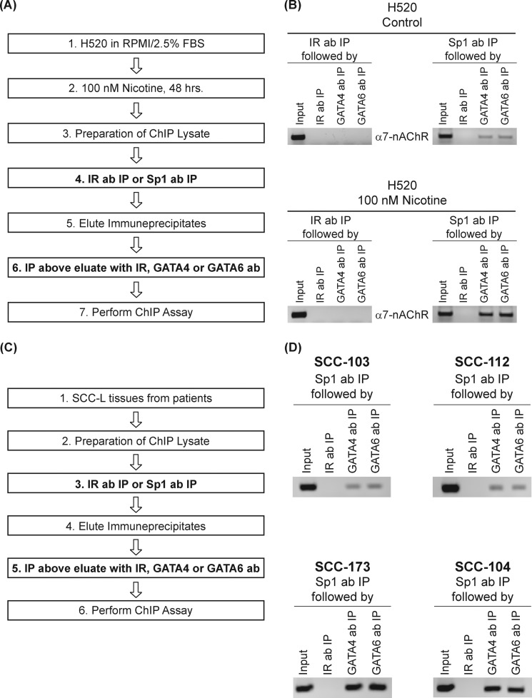
ChIP-re-ChIP assays reveal increased Sp1-GATA4 and Sp1-GATA6 complexes on the human α7-nAChR promoter in nicotine-treated H520 cells and human SCC-Ls. A, a flowchart describing the sequential steps involved in ChIP-re-ChIP experiment in H520 cells. B, untreated H520 control cells had very low amounts of Sp1-GATA4 and Sp1-GATA6 complexes on the α7-nAChR promoter (top right panel). The onset of nicotine treatment led to robust amounts of Sp1-GATA4 and Sp1-GATA6 complexes on the α7-nAChR promoter (bottom right panel). The antibodies used the second round of chromatin IP, namely GATA4 polyclonal antibody and GATA6 polyclonal antibody, are indicated vertically next to the individual lanes. C, a flowchart describing the sequential steps involved in ChIP-re-ChIP experiment in human SCC-L tumors (isolated from patients). D, SCC-103 and SCC- 112 human SCC-L tumors had lower levels of Sp1-GATA4 and Sp1-GATA6 complexes bound to the α7-nAChR promoter (top panels) compared with SCC-173 and SCC-104 human SCC-L tumors (bottom panels). The input DNA lane represents one-fifth of the precleared chromatin used in each ChIP reaction. The figure is representative of two independent experiments.

