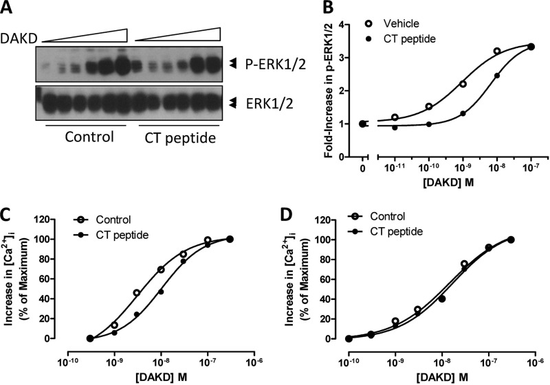
FIGURE 8.
Disruption of CPM/B1R interaction with CT peptide decreases B1R-dependent signaling. HEK cells stably expressing B1R and CPM were incubated without or with 50 μm CT peptide for 10 min. Cells were stimulated with various concentrations of B1R agonist DAKD for 5 min, and total and phospho-ERK1/2 were determined by Western blotting (A). Bands were quantified by densitometry, and phospho-ERK1/2 values were normalized to the density of total ERK1/2 (B). HEK cells stably co-expressing B1R and CPM (C) or B1R alone (D) were incubated without or with 50 μm CT peptide for 10 min after Fura-2/AM loading, and B1R-dependent increase in [Ca2+]i was recorded in response to various concentrations of DAKD. The data are representative of three experiments.

