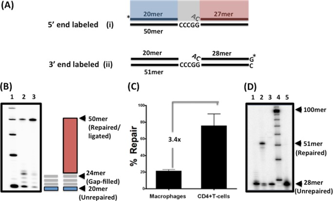
FIGURE 1.
In vitro DNA gap repair activity of nuclear extracts from human primary activated CD4+ T cells and MDMs. A, 32P-5′-end-labeled (substrate i) or 32P-3′-end-labeled (substrate ii) gapped DNA substrate was used in this study as described previously (6). Substrate i consists of a 20-mer upstream primer (blue) and a 27-mer downstream primer (red), which are annealed to a 50-mer template, creating a four-nucleotide gap (gray) with a 5′-end flap of the downstream primer. B, nuclear extracts normalized to 2 μg/μl total protein from either macrophages (lane 2) or CD4+ T cells (lane 3) from three donors (data from one donor are shown) were incubated with the 5′-end gapped substrate for 30 min at 37 °C. Reactions were quenched with 40 mm EDTA and subjected to 14% urea-PAGE. Gap filling generated a 24-mer product, followed by a fully repaired 50-mer product as indicated. A 10-mer ladder was run in lane 1. C, repair capacity comparison of macrophages and activated CD4+ T cells from three donors. The percent of the fully repaired 50-mer product in the total loading in each lane was quantified by densitometry, and the -fold difference between CD4+ T cells and macrophages was calculated. D, substrate ii containing the 3′-end-labeled 28-mer downstream primer was incubated with nuclear extracts normalized to 2 μg/μl total protein from macrophages in the presence (lane 2) or absence (lane 3) of 250 μm dNTPs for 30 min at 37 °C. Lane 1 is the control reaction without nuclear extract. Reactions were quenched with 40 mm EDTA and subjected to 14% urea-PAGE. The unrepaired 28-mer downstream primer and the fully repaired 51-mer product are indicated. A 10-mer ladder (lane 4) and the 28-mer control downstream primer (lane 5) are also shown.

