
FIGURE 5.
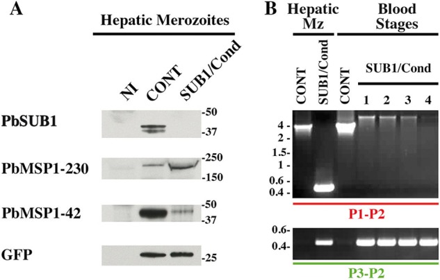
SUB1/Cond hepatic merozoites display a default of MSP1 maturation and are unable to establish a blood parasitemia. A, protein extracts of CONT and SUB1/Cond hepatic merozoites mechanically released from infected HepG2 cultures 60 hpi were probed with the 1D11 anti-PbSUB1 mAb and the rabbit anti-PbMSP1 polyclonal serum. Anti-GFP antibodies were used as a parasite loading control. Molecular mass markers are in kilodaltons. B, PCR genotyping of the sub1 locus using the P1-P2 and P3-P2 primers (Fig. 2A). gDNA was prepared from SUB1/Cond or CONT hepatic merozoites (Mz) and blood stage parasites collected from mice infected by 50,000 hepatic CONT or SUB1/Cond merozoites, corresponding to experiment 3 presented in Table 2. PCR analysis of one representative CONT gDNA is shown, whereas lanes 1–4 correspond to the analysis of the gDNA prepared from the four mice that developed a blood parasitemia following the injection of SUB1/Cond hepatic merozoites (Table 2). The SUB1/Cond genotype of these parasites was confirmed by amplification of the 0.43-kb fragment by the P3-P2 primers. However, no parasites with an excised Pbsub1 locus could be detected, as shown by the failure to amplify the Pbsub1 excised specific fragment of 0.4 kb using the P1-P2 primers, whereas the full-length Pbsub1 non-excised locus is detected, as shown by the amplification of the 5.5-kb fragment (Fig. 2A). Molecular mass markers are in kilobase pairs.