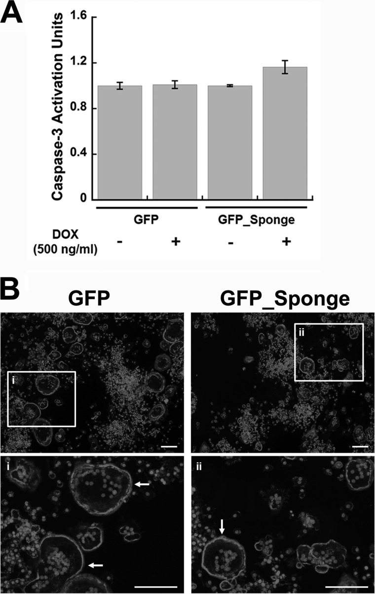
FIGURE 7.
Inhibition of miR-29 does not affect apoptosis of mature osteoclasts, or actin ring formation. A, GFP and GFP_Sponge RAW264.7 cells were differentiated for 2 days with RANKL (30 ng/ml). The expression of the transgene was induced by addition of DOX (500 ng/ml) at day 3. Caspase-3 activity in mature osteoclast cultures was quantified after 4 days of differentiation (n = 6). B, GFP and GFP_Sponge RAW264.7 cells were cultured on glass chamber slides for 4 days in the presence of DOX (500 ng/ml) and RANKL (30 ng/ml). Actin ring formation was evaluated by phalloidin staining, and nuclei were visualized with DAPI reagent. Representative images were captured using 5× magnification. Boxed regions i and ii are visualized at 20× magnification. Actin rings are indicated by arrows. Scale bar, 100 μm.

