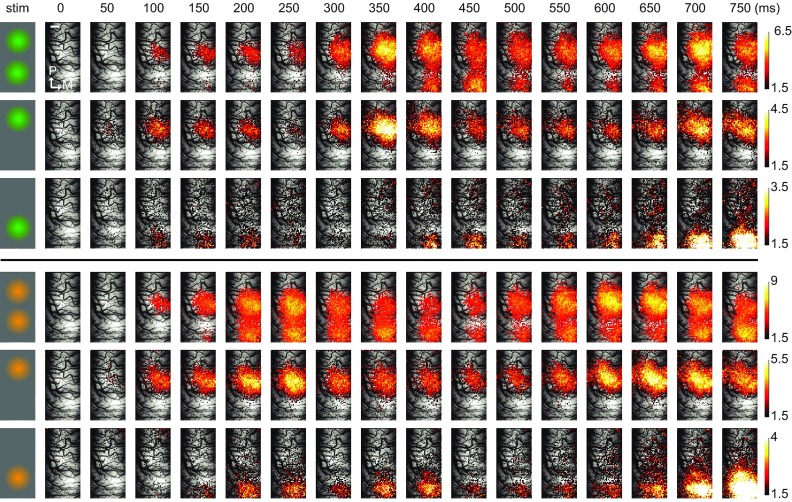
Figure 2. Dynamics of cortical activity evoked by locally presented natural stimuli.
( A) Spatiotemporal activity patterns produced by locally presented natural movies is shown overlaid on the vascular image. Leftmost column schematically shows the corresponding stimulus condition and the movie used ( orange and green). Each single frame represents the average activity across trials (n = 35) within 50 ms of non-overlapping segments of recorded data. The cortical representations of these stimuli were located along the antero-posterior axis, reflecting the vertical positioning of the movie patches in the visual field. Different spatial extents and peak values of cortical activation were observed along the presentation duration at different frames. Please note that different color-scales are used for different conditions in order to emphasize the detailed spatial structure. Upper color scale is clipped to 3.3 standard deviations. Only the most active pixels corresponding to highest 25 th percentile are shown. For a video presentation please refer to the Video S2. Scale bar (shown in the first row in the frame zero) = 1mm.

