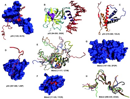
Figure 14. Structural characterization of human proteins involved in the p53-mediated apoptotic signaling pathways: group IV.

A. Residues 1–93 of p53 (red ribbon) in a complex with the bromodomain of CREB-binding protein (blue surface) (PDB ID: 2LY4); B. Residues 94–293 of p53 in a complex with DNA (red bonds) (PDB ID: 3IGK); C. Residues 319–360 of p53 in a homotetrameric complex (PDB ID: 1OLH); D. Residues 367–386 of p53 (red ribbon) in a complex with the bromodomain of CREB-binding protein (blue surface) (PDB ID: 1JSP); E. Residues 1–117 of MDM2 (PDB ID: 1Z1M); F. Residues 17–125 of MDM2 (blue surface) in a complex with the transactivation domain of p53 (residues 15–29, red ribbon) (PDB ID: 1YCR); G. Residues 147–150 of Mdm2 (red ribbon) in a complex with the N-terminal domain of HAUSP/USP7 (blue surface) (PDB ID: 2FOP); H. Residues 290–335 of Mdm2 (PDB ID: 2C6A).
