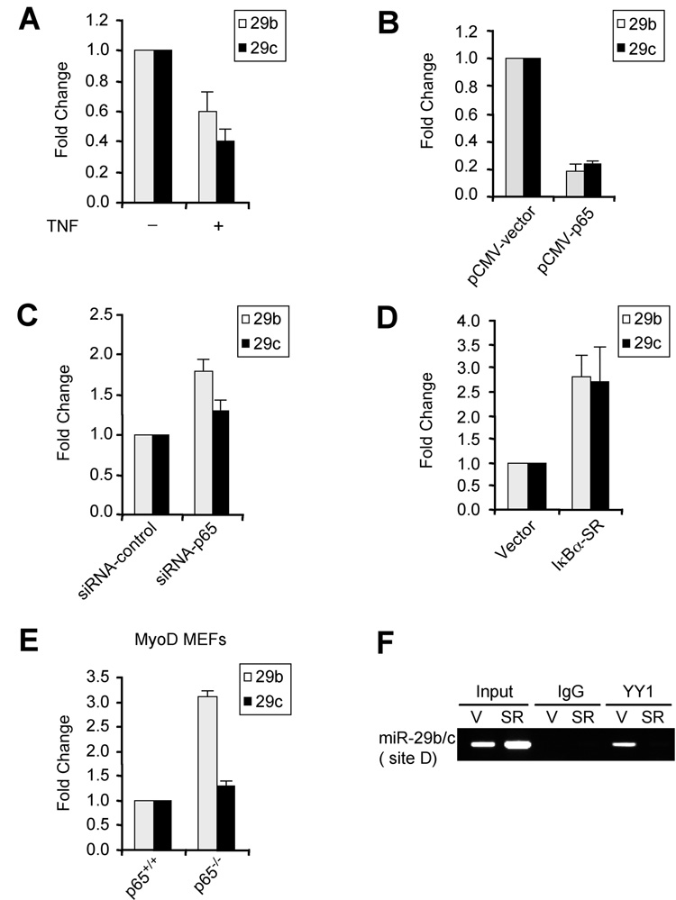
Figure 2. NF-κB negatively regulates miR-29b/c.
(A) C2C12 cells were treated with TNFα and miR-29 was measured by qRT-PCR normalized to U6. Fold changes are shown with respect to vector cells where miR-29 levels were set to a value of 1. (B) MB were transfected with vector or a p65 plasmid and miR-29b/c levels were measured 48h post-transfection. (C) MB were transfected with either vector or p65 siRNA oligos and miR-29 expression was then measured as in (B). (D) MiR-29b/c were measured in MB stably expressing vector or IκBα-SR. Fold changes are shown with respect to vector cells, which were set to a value of 1. (E) MyoD was stably expressed in p65+/+ or p65−/− mouse embryonic fibroblasts (Bakkar et al., 2008) and qRT-PCR was performed for miR-29b and miR-29c. (F) ChIPs with YY1 or control IgG were performed on chromatins derived from either vector control (V) or Iκ Bα-SR (SR) expressing MB. Primers specific to site D were used for the PCR amplification. Total inputs are indicated.

