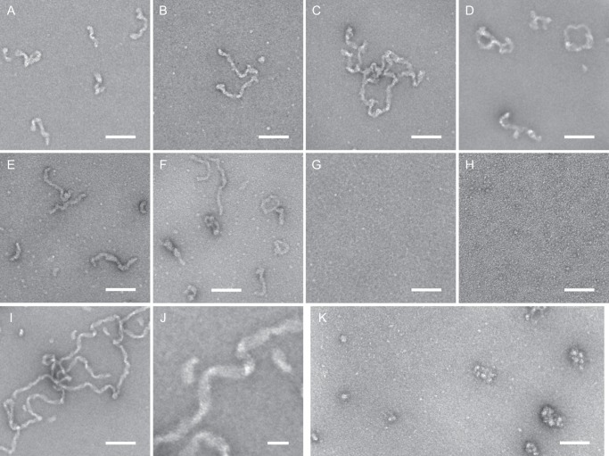
FIGURE 5.
Transmission electron micrographs of ameloblastin and its mutant variants. Purified AMBN (A–D), AMBN-Nterm (E and F), AMBN-Cterm (G), AMBNΔ36–72 (H), S36–72-AMBN-Cterm (I and J), and AMEL (K) were applied onto glow discharge-activated carbon coated grids and negatively stained with 2% uranyl acetate. Samples were examined at a magnification of 64,000×. J shows a 2.84-fold digitally magnified detail of the ribbon-like structure of the S36–72-AMBN-Cterm protein filament presented in I. Scale bars, 100 nm (A–I and K), or 25 nm (J).

