
FIGURE 2.
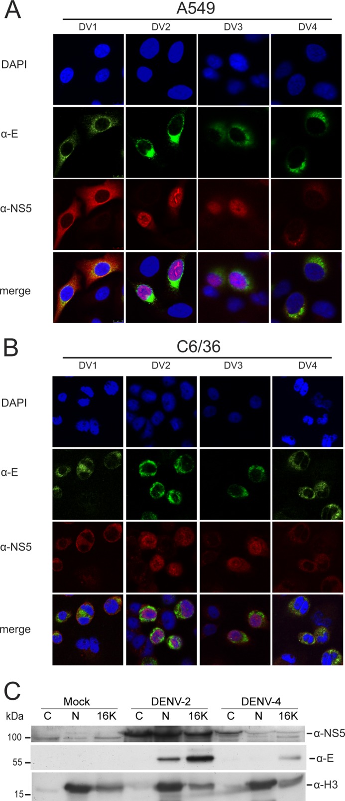
Subcellular localization of DENV-1 to -4 NS5 during viral infection. CLSM images of A549 cells (A) and insect C6/36 cells (B) infected with DENV-1, -2, -3 and -4 at a multiplicity of infection of 2.0 and immunostained using anti-E (α-E) and anti-NS5 (α-NS5) antibodies at 30 h postinfection. In each panel, a selection of infected (as evidenced by anti-E staining) and non-infected cells are shown as a control for nonspecific staining by the anti-NS5 antibody. Nuclear DNA was stained with DAPI. C, Western blot analysis of cytoplasmic (C), nuclear (N), and cytoplasmic membrane (16K) fractions prepared from Huh-7 cells at 30 h postinfection with DENV-2 or -4 (multiplicity of infection of 3) or mock-infected. 10 μg of protein from each cellular fraction was analyzed for the presence of the DENV NS5 and E proteins and histone H3 using specific antibodies by Western blotting. Histone H3 was used as a nuclear marker. The positions of relevant molecular mass markers are shown in kDa.