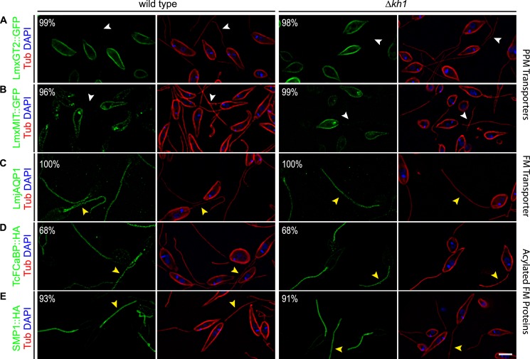
FIGURE 7.
Targeting of other membrane proteins in Δkh1 mutant cells. Immunofluorescence of PPM proteins and other proteins known to specifically target to the FM. Green is GFP, HA, or LmjAQP1; red is tubulin; blue is DAPI. White arrowheads indicate lack of flagellar localization. Yellow arrowheads indicate flagellar localization. Percentage shown in panels represent the fraction of cells with localization as shown. Scale bar = 3 μm.

