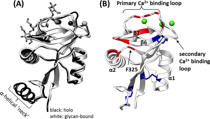
FIGURE 1.

Insight from current crystal structures. A, comparison of holo-structure (Protein Data Bank code 1XPH; black) and (GlcNAc)2Man3-bound (1K9J; white) crystal structure. Ca2+ ions are represented by spheres. All published crystal structures adopt nearly identical conformations, suggesting that the DC-SIGNR CRD adopts the same conformation during crystal formation with or without glycan. B, residues that form direct contacts/bonds with the glycan in the structure of the (GlcNAc)2Man3·CDR complex (1K9J; ligand not shown) are highlighted in red, disulfide bonds are shown in blue, and bound calcium ions are shown in green.
