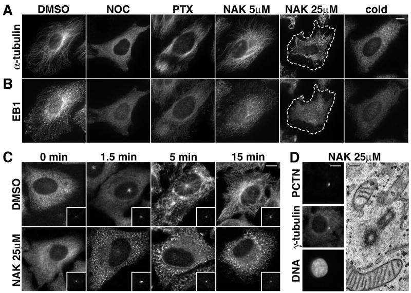
Figure 3. Nakiterpiosin suppresses microtubule dynamics in interphase cells.
A and B, HeLa cells were treated for 2 h with DMSO, 5 μg/ml nocodazole (NOC), 1 μM paclitaxel (PTX), 5 or 25 μM nakiterpiosin (NAK), or cooled on ice for 30 min (cold). The cells were then fixed and labeled for α-tubulin (A) and EB1 (B). Bar, 10 μm.
A, In 25 μM NAK microtubules no longer originated from the centrosomes and became shorter (The cell border is outlined with dash lines). 5 μM NAK and DMSO showed little or no effect. B, EB1 was concentrated at the tips of microtubules in DMSO but not in 25 μM NAK, suggesting NAK suppresses microtubule dynamics.
C, Centrosome-mediated microtubule regrowth is inhibited by 25 μM NAK. HeLa cells were cooled on ice for 30 min to depolymerize microtubules and incubated on ice for 30 min with DMSO or 25 μM NAK (0 min). The cells were then warmed to 37°C, fixed at the indicated time points and double-labeled for α-tubulin and pericentrin (insets). Bar, 10 μm.
D, Nakiterpiosin does not alter centrosome organization. HeLa cells treated for 2 h with 25 μM NAK were either immunolabeled for pericentrin (PCTN), γ-tubulin and DNA (left panels, bar, 10μm) or analyzed by EM (right panel, bar, 200 nm).

