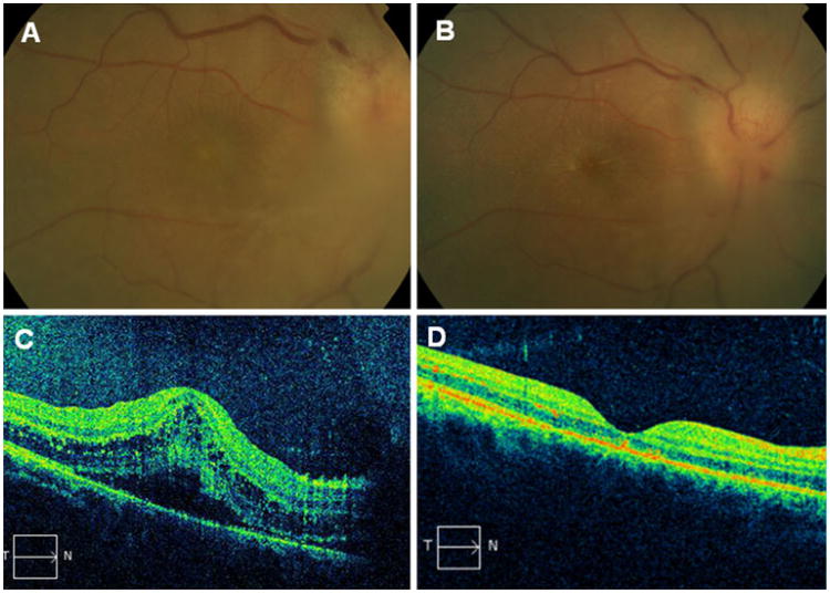
Fig. 1.
a Right eye color fundus photograph at presentation shows inflammatory infiltrate over the edematous optic nerve. b Right eye color fundus photograph 1 week later shows optic disc edema with macular star exudates. c OCT scan of the right eye at presentation shows serous macular detachment with intraretinal cystic changes. d OCT scan 1 month later shows complete resolution

