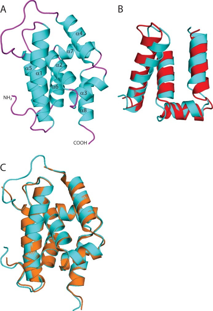
FIGURE 2.

Structure of the ClpC1 N-terminal domain. A, overview of the structure of M. tuberculosis ClpC1 NTD, shown in schematic representation with the α-helices (cyan) and loops (magenta). The amino and carboxyl termini are indicated and the α-helices are labeled. B, structural alignment of ClpC1 residues 4–68 (cyan) with residues 79–143 (red; r.m.s. deviation 2.035). C, structural alignment of M. tuberculosis ClpC1 NTD (cyan) with B. subtilis ClpC NTD (orange; r.m.s. deviation 0.48).
