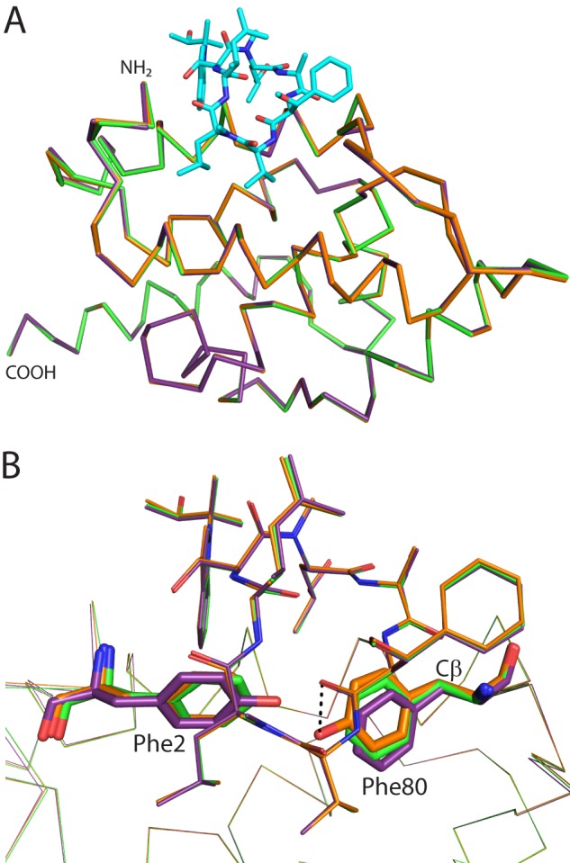
FIGURE 4.

Comparison of CymA binding to WT ClpC1 versus F2Y or F80Y mutants. A, backbone alignment of the structures of WT (green), F2Y (purple), and F80Y (orange) ClpC1 NTD bound to CymA. CymA is shown as cyan sticks and ClpC1 as ribbons. The amino and carboxyl termini are indicated. B, the same orientation as in A with CymA from each structure shown as lines and ClpC1 shown as ribbons. Phe2 and Phe80 (or their Tyr mutants) are shown as sticks. CymA and ClpC1 are colored: WT, green; F2Y, purple; F80Y, orange. A potential hydrogen bond interaction between Tyr80 and a carbonyl of CymA is shown as a dashed line. The Cβ atom discussed in the text is indicated.
