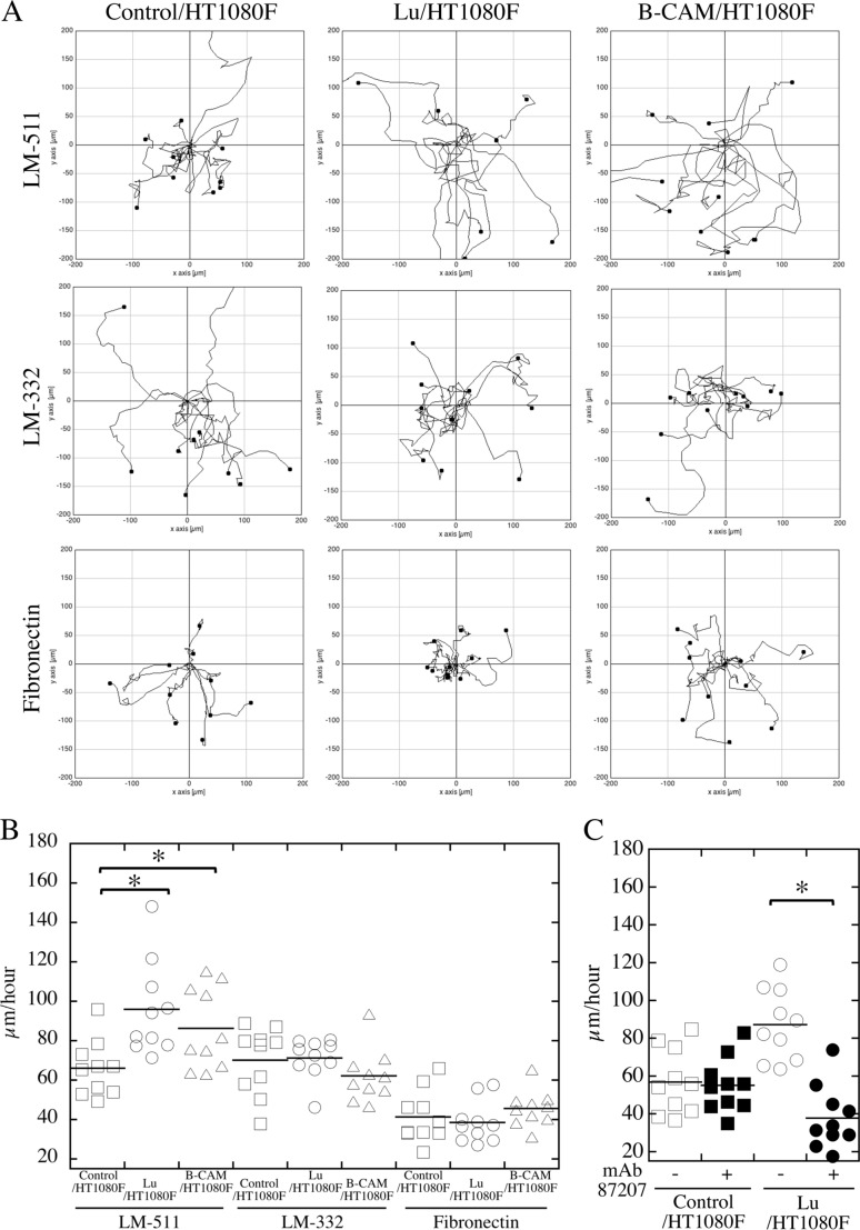
FIGURE 5.
Pattern and velocity of cell migration. The indicated cells were plated on dishes coated with LM-511 (0.8 nm, top panels), LM-332 (0.8 nm, center panels), or fibronectin (40 nm, bottom panels). Cell movements were monitored by time-lapse video microscopy. A, representative paths of cell movements on each proteins tracked at 10-min intervals over a span of 4 h. B, quantification of cell motility as evaluated by velocity (micrometers/hour) and determined using ImageJ software as described under “Experimental procedures.” *, p < 0.01 by Dunnett's multiple comparison test. C, mAb 87207 inhibited migration of Lu/HT1080F cells on LM-511. After cell adhesion to substratum, mAb 87207 was added to culture medium. After another hour, cell movements were monitored for 4 h. Cell motility was evaluated by velocity as described above.

